1
2
3
4
5
6
7
8 |
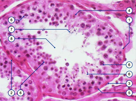
Basal lamina (membrane) (not recognizable)
Myofibroblast
Fibrocyte
Sertoli's cell
Spermatogonia
Various stages of the germ cells during spermatogenesis
Spermatozoon
Lumen |
|
|
|
Fig. 10
Histological preparation of a section through a convoluted seminiferous tubule in an adult. Outside its basal lamina a layer of myofibroblasts and fibrocytes surround the tubule. The germinal epithelium lies on the tubule wall. One can recognize the spermatogonia sitting on the basal lamina.
The nuclei of the Sertoli's sustentacular cells have a rarified chromatin and the nuclei with clear nucleolus that are often oriented perpendicular to the basal lamina. The overall picture, though, is dominated by the cells occupied with spermatogenesis.
|