|

|
|
|
|
T cells mature out of stem cells produced in the bone marrow and transported into the thymus. They emigrate out of the thymus, colonizing the secondary lymph organs, and are active there as immunocompetent cells for the defense of the body against infections. The thymus derives from the foregut out of the 3rd and 4th pharyngeal pouches. Its stroma arises out of epithelial cells of ectodermal and also endodermal origins.
|
|
|
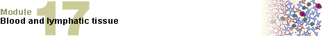
| Fig. 8 - Embryo in stage 13 (ca. 32 days) |
|
Legend |
1
2
3
4
5
6
7
8 |
Thyroid and foramen cecum
1st pharyngeal arch (mandibular arch)
Aorta dorsalis (paired)
Neural tube
Anlage of the thymus
1st pharyngeal fold (anlage of the external ear)
2nd pharyngeal fold
Entrance to the cervical sinus |
|
|
|
Fig. 8
In order to better show the relationships in the foregut, a cross-
section at the level of the pharyngeal arches has been made. One looks at the ventral part of the mouth/throat cavity. The externally visible pharyngeal folds correspond internally to the pharyngeal pouches.
|
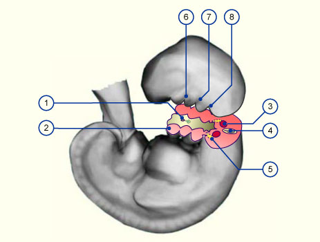
| Fig. 9 - Thymus development |
|
Legend |
1
2
3
4
5
6
7
8
9
10 |
Mandibular arch
Hyoid arch
Entrance to the cervical sinus
3rd pharyngeal pouch
4th pharyngeal pouch
Foramen cecum
Thyroid
Cervical sinus
Thymus (part of the 3rd pharyngeal pouch)
Thymus (part of the 4th pharyngeal pouch) |
|
|
|
Fig.9
View of the cross-
section of Fig. 8
|
|
More info
|
|
In the foregut region the relationships are complicated (overview of the most important derivatives of the foregut).
|
|
|
|
|
|
Up to the sixth week, the thymus is purely epithelial. In the next weeks it becomes dented by an in-growing mesenchymal septal. In this way arise pseudo-lobules that communicate with each other in the center. The mesenchymal anlage material further forms the numerous, partially widened, perivascular spaces that are important for the cell transport into and out of the thymus. The fully developed organ consists of the thymus-epithelial tissue, responsible for the maturation of the T lymphocytes, and the perivascular spaces.
Despite being tightly woven together they are separated from one another by a layer of flat epithelial cells with a basal lamina, the thymus-blood barrier. The anlage material for the non-epithelial stromal cells (precursors of the macrophages) and the free cells of the thymus (prethymic stem cells of the T lymphocytes) derive from embryonic blood formation centers (liver, spleen, bone marrow). They appear in the thymus after the 9th week and initially colonize it relatively uniformly. The thymus (histological picture) acquires its functioning ability only when the cortex and medulla have differentiated. The first indications of this differentiation in a fetus occur after ca. 12 weeks and it is completed at ca. 4 months. With this differentiation the young thymocytes settle into the cortex region.
|
|
|
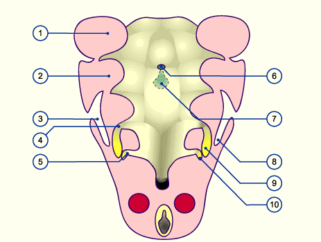
| Fig. 10 - Construction of the thymus (without T cells) |
|
Legend |
1
2
3
4
5
6
7
8
9
A
B |
Thymus capsule
Thymic nurse cells
Connective tissue septa with blood vessels
Subcapsular epithelium (blood-thymus barrier)
Cortical epithelial cells
Medullar epithelial cells
Dendritic cells (from the bone marrow)
Hassall's corpuscle (histological picture)
Macrophages (from the bone marrow)
Cortex
Medulla |
|
|
|
Fig. 10
In the cortex, the lymphocyte precursors are thickly packed together and are closely associated with the "thymic nurse cells". Their differentiation takes place through migration into the medullar region, where the cells are more loosely arranged. Moreover, the thymus consists of cortical and medullar epithelial cells. A series of smaller epithelial cells that form the blood-thymus barrier surround the individual lobes and the capillaries (not shown). A large number of macrophages also settle in the thymus that, like the thymocytes, derive from liver and bone marrow hematopoietic stem cells.
|
|
More info
|
|
The thymus stroma plays a decisive role in the further development of the thymocytes into T cells. The epithelial cells of the thymus medulla secrete chemokines (MDC = macrophage-derived chemokines) that chemotactically attract the immature thymocytes, i.e., the thymocytes express binding sites that are highly specific for these MDC. These MDC are probably responsible for the maturation of the thymocytes (interactive diagram, 41 kb) during their migration from the cortex into the medulla. (5)
|
|
|
|
|
|
|