|

|
|
|
21.3 Differentiation of the gonads
|
|
|
Gonads: indifferent stage
|
|
|
|
The gonads arise from two very different kinds of cells that originate in the embryo:·
- The primordial germ cells (PGC) will form the gametes (sperm cells and oocytes). These cells come from the ectoderm, but they separate themselves from it at a very early stage in the development.
- The somatic cells with nourishing functions surround the primordial germ cells and form the somatic gonadal blastema. In the testes the supporting cells (Sertoli) and the interstitial cells (Leydig) are involved, in the ovary the follicle cells and the theca cells.
Their origin is still a matter of discussion, however, whereby three possible sources come into question: mesonephros, local mesenchyma, as well as superficial epithelial cells (coelomic epithelium).
|
|
|
Fig. 4 - Migration of the PGC
4th – 6th Week |
|
Fig. 5 - Migration of the PGC
4th – 6th Week |
|
Legend |
1
2
3
4
5
6
7
8
9
|
PGC
Allantois
Cloacal membrane
Epiblast
Pharyngeal membrane
Heart anlage
Umbilical vesicle (yolk sac) Endoderm
Mesoderm |
|
|
|
1
2
3
4
5
6
7 |
Rectum
Omphalomesenteric duct
Allantois
Nephrogenic cord (pink)
Gonadal ridge (green)
PGC
Heart anlage |
|
|
|
Fig. 4
After their emigration into the umbilical vesicle the PGC enter again into the embryo and thence into the intestinal wall.
Fig. 5
From the intestine they colonize the gonadal ridge in that they emigrate there via the mesentery.
|
|
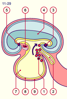
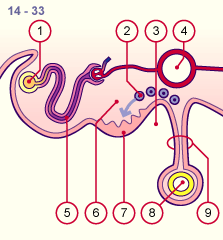
Like the nephrogenic cord the gonadal ridge extends from the heart region to the location near the cloaca. In the time between the 4th and 6th week  11-14 11-14 the middle section of this gonadal ridge develops into a gonad anlage in that cells of the coelomic epithelium proliferate there. The immigrated PGC 14 penetrate into this thickened zone of the coelomic epithelium. The indifferent gonads thus assemble themselves from cells of various origins, whereby, as a result, the primordial germ cells and the local somatic blastema influence each other reciprocally (13). the middle section of this gonadal ridge develops into a gonad anlage in that cells of the coelomic epithelium proliferate there. The immigrated PGC 14 penetrate into this thickened zone of the coelomic epithelium. The indifferent gonads thus assemble themselves from cells of various origins, whereby, as a result, the primordial germ cells and the local somatic blastema influence each other reciprocally (13).
|
|
|
Definition
|
|
Blastema: indifferent collection of embryonic cells
|
|
|
|
The local coelomic mesenchyma underneath also multiplies. The coelomic epithelium, which now becomes multi-layered, loses for now its basal membrane. Gonadal cords arise that surround the PGC and extend into the depths. One knows that - in the male - mesonephric cells are also involved, but it is unknown whether in females the cells immigrate as far as the gonadal ridge (14).
Up to the 6th week male and female gonads cannot be distinguished. The gonadal cords and the PGC can be found both in the cortical as well as in the medullar zones of the future gonads.
|
|
|
Fig. 6 - Indifferent gonads
Stage 14, ca. 33 days |
|
Fig. 7 - Indifferent gonads
stage 17, ca. 41 days |
|
Legend |
1
2
3
4
5
6
7
8
9 |
Mesonephric duct (Wolff)
PGC
Peritoneal cavity
Aorta
Mesonephric tubule
Local coelomic mesenchyme
Thickened coelomic epithelium
Intestine
Mesentery |
|
|
|
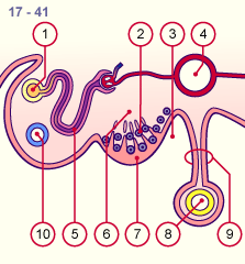
1
2
3
4
5
6
7
8
9
10 |
Mesonephric duct (Wolff)
PGC
Peritoneal cavity
Aorta
Mesonephric tubule
Local coelomic mesenchyme
Thickend coelomic epithelium
Intestine
Mesentery
Anlage of the paramesonephric duct (Müller) |
|
|
|
Fig. 6
The PGC immigrate into the genital ridge. At this time, the mesonephros structures develop in the mesonephric ridge.
Fig. 7
Cells of the thickened coelomic epithelium leave the cellular envelope and migrate into the depths where they form the gonadal cords with the PGC and the local mesenchyma. The anlage of the paramesonephric duct (Müller) is now formed.
|
Fig. 8 - Early development in the female
stage 18, ca. 49 - 51 days |
|
Fig. 9 - Early development in a male
stage 18, ca. 44 - 49 days |
|
Legend |
1
2
3
4
5
6
7
8
9
10
11 |
Mesonephric duct (Wolff)
PGC
Peritoneal cavity
Aorta
Mesonephric tubule
Degenerated gonadal cords
Thickened coelomic epithelium
Intestine
Mesentery
Anlage of the paramesonephric
duct (Müller)
Atrophying mesonephric nephron |
|
|
|
1
2
3
4
5
6
7
8
9
10
11 |
Mesonephric duct (Wolff)
PGC
Peritoneal cavity
Aorta
Mesonephric tubule
Gonadal cords
Coelomic epithelium
Intestine
Mesentery
Anlage of the paramesonephric
duct (Müller)
Mesonephric nephron |
|
|
|
Fig. 8
In females, the gonadal cords retain their connection to surface epithelium. The PGC are now found mainly in the cortex. Only a few of the gonadal cords reach the medulla.
Fig. 9
In males the testicular cords, consisting of thickly packed prospermatogonia and somatic cells, develop out of the gonadal cords. These come from the coelomic epithelium (Sertoli's supporting cells), the local mesenchyma and the mesonephros. The testicular cords extend deeply into the region of the future testicles.
|
|
|